![]()
![]()
The ability to detect the odor of molecules is probably the oldest of senses for life on earth. The most primitive single celled organisms living in water have the ability to taste the presence of food around them. While we often distinguish our sense of smell from our sense of taste, the distinction is largely a matter of location rather than fundamental differences. Similar chemical structures and mechanisms are used by all of our senses as well as in other cells types, even those of primitive organisms. This suggesting a history of diverging evolution rather than independent development of each sensory system.
The chemistry of taste and smell may be simplest on the molecular level. But human experiences are at a scale many orders of magnitude larger (by 105 to 1011). It is easier to understand mechanisms not far removed from our experiences. So because it is easier for us to understand, the system we use for hearing is considered first, next the senses of touch, followed by taste and smell.
Based on the many historical examples where early scientific explanations were later modified by improved understanding, some of the following explanations of our senses may also be refined as experiments provide additional evidence. With that caveat, we proceed:
 Hearing is provided by the organ we label our
 ears.
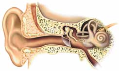
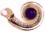
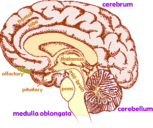
(As is always the case, our label provides no explanation or understanding, but just gives a handle to link understanding together, here to connect with your early lessons of what ears look like and where they are located. Such is the function and value of all labels including the many which follow.) Sound vibrations transmitted through air are funneled into our heads by our outer ear (←diagram left). At the end of a short, protective canal, the vibrating air causes a delicate membrane to vibrate. Three tiny bones act as levers transmitting vibrations from the back side of this outer membrane to a second membrane which caps a coiled cone-shaped, fluid-filled inner chamber (image above right→). Ebb and flow of this fluid cause the sway of a field of tiny hair-like cilia (at right & further magnified below right), each with a nerve attached at its base.
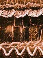
The sound vibrations produce shearing forces which bend each cilia. Just like we can feel the motion when we move hair elsewhere on our bodies, when cilia motion is detected the nerves attached to these cilia send electrical signals to the brain. This bending opens tiny channels in the cell membrane, allowing positively charged Potassium ions, K+, to flow out of the cell, depolarizing the small electrical potential normally existing across the membrane of the cell. This initiates a electrical nerve signal flowing along the outer membrane of the nerve. This signal is pre-processes by neurons (nerve cells) of the spiral ganglia which then forward a signal of detected sound off to the brain via the eighth cranial nerve. Every generic rod has a natural vibrational frequency depending on its length and mass. Like pumping a child on a swing, a cilia will gain the greatest motion when pumped by a vibration that matches the natural frequency of swing. Different length cilia move more depending on the frequency of the vibration. So the brain interprets the particular frequency (pitch) of sound vibration by which nerves report their cilia are moving the most. Loudness is presumably interpreted by the extent of the cilia motion and original strength of electrical signal.
All living cells have clusters of protein molecules imbedded in their cell membranes which act as gateways into and out of each cell. One such protein consumes energy supplied in the form of the molecule ATP to actively pump Potassium ions, K+, into the cell and smaller Sodium ions, Na+, out. The difference between Potassium and Sodium ion attractions for electrons creates a small electrical potential (measurable as Voltage) across the cell membrane. The part of nerve cells which function as sensors (called receptors) have specialized protein gateways which upon stimulation allow ions to spontaneously leak back across the cell membrane to regions of lower concentration, thereby reducing and briefly reversing this electrical potential. This electrical discharge caused by ions flooding through the membrane, then flowing parallel to the membrane surface surface triggers the opening of neighboring gateways. This creates an avalanche of electrical discharge which, crucial to all the senses, travels like a wave across the nerve's outer surface, transmitting a message a little like voltage pulses are used to carry information in electronic devices. The protein ion pumps subsequently re-establish the concentration gradients of Potassium and Sodium ions across the cell's membrane, restoring the electrical potential, thus preparing the nerve for another message transmission.
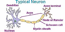
Unlike many cells which have approximately spherical shape maximizing their content compared to their required outer surface membrane, most neurons have a long thread-like out-stretched tentacle called an axon, sometimes a meter or more long. And a second difference, while most cells are surrounded by water containing ions which quickly dissipates any electrical discharge, axons are often buried inside by a chain of specialized Schwann cells which provide a water insoluble, fatty myelin sheath with a high dielectric constant, which insulates the discharge from the effect of ions in nearby water. As the voltage spike passes, its electric field exerts electrical force on any nearby ion. This shares a portion of the voltage spike's energy, fading the signal's strength. Such interaction also slows the speed of message transmission along the axon. By keeping ions away from the axon, the myelin helps speed the signal transmission and avoid loss of signal. Even with the myelin shielding, the top speed of nerve pulses is in the order of hundreds of m/s, far slower than electrical signals traveling the speed of light along metal conductors.
The skin contains a variety of receptors (sensor portion of nerves) which provide our senses of touch or pressure, temperature (warm or cold), and pain (including itch and tickle). The same system also provides sensations of muscle movement and joint position including posture, movement, and facial expression. It also provides sensory information from internal organs, such as stomach aches. We use these senses to interpret information such as shape, softness, texture, and vibration.
The mechanism used by the senses of touch is similar to that described for hearing. The receptor is either a free nerve ending
or a nerve ending embedded in a specialised capsule. A receptor is activated by movement or pressure (mechanoreceptor), reaction with a chemical substance (chemoreceptor) and/or temperature. In each case, the stimulus causes depolarisation of the nerve ending causing a wave of changing electrical potential (Voltage spike) to be propagated across the outer membrane of the neuron. This signal generally travels along the axon towards the spinal cord.
Animals such as ourselves use these electrical discharges to transmit messages along nerve membranes the often significant distances from sense organs to the brain, and motor signals back to distant muscle tissue.
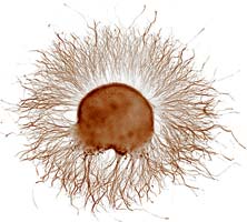
The typical sensory nerve pathway is three neurons long. The first neuron always has its cell body in the dorsal root ganglion of the spinal nerve. (←Image at left shows axons growing from such a dorsal ganglion of a developing chicken embryo.) The second neuron has its cell body either in the spinal cord or in the brainstem (medulla oblongata) with the axon terminal end in the thalamus, or the cerebellum. In the case of touch and certain types of pain, the third neuron has its cell body in the thalamus and ends near the top of the brain in the cerebrum.
Now that we are investigating processes where one can't easily experiment in a beaker or otherwise outside of a living creature, one might asks how to proceed? One answer: Carefully! Whenever working with a living being, particularly with a person, there is a need for more care and considerable concern for the welfare of the person involved. And if you decide to do even trivial experiments on yourself, additional hazards should be considered! So proceed with care and prior planning.
This is a particularly appropriate time to ask the questionWhy experiment at all?The writings of Confucius dating back 2500 years contain the basis for the answer:
Ideally readers should formulate their own investigations to match what they wish to better understand. But many readers have had little practice and are often unclear how to proceed. So each investigation generally contains some suggested possibilities to provide a basis at experimenting. Unlike many efforts at science instruction, the goal here is NOT to provide a cookbook telling the reader every detail of what to do, but rather provide some prudent guidance, allowing the reader to ponder, and occasionally search down dead-end paths. If the reader takes the time and does such investigation, the results will usually be superior understanding of the topic under study as well as a keen awareness of the nature of knowledge and various scientific skills. Those are the intended goals of the author and hopefully also of you the reader!
Communicating technical information such as observations and findings is a skill used by scientists but useful for most others. If you need course credit, use your observations in your journal to construct a formal report.
![]()
to next investigation
to Biochemistry menu
to ie-Chemistry menu
to site menu