![]()
![]()
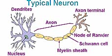 Earlier investigations explained how nerve receptors found in humans and many other organisms sense aspects of their environment (touching & hearing and tasting & smelling) and conditions inside their body (pain). This information is converted to electrical signals (described in pain and nerve messaging) in the form of a rapid reversal of electrical potential (a Voltage spike) across the cell membrane of neurons (nerve cells). Powered by energy stored in the normal segregation between ions outside and inside the cell, the nerve signal travels along the long thin axon over distances of up to several meters in large animals. Eventually the signal reaches the brain for analysis, response, and memory.
Many earlier peoples believed that the heart was the seat of human intelligence. (We still memorize by heart, and express our devotion with Valentine's hearts.) But it is now well established that the brain is the control center of the central nervous system, responsible for such functions as reason and intelligence, including cognition, perception, attention, memory and emotion. A brain also controls positions of movable parts and movement of the entire creature. It makes possible learning. The brain also performs a variety of automatic functions such as coordination of walking and sensory systems, and internal functions such as regulating blood pressure, fluid content, and body temperature.
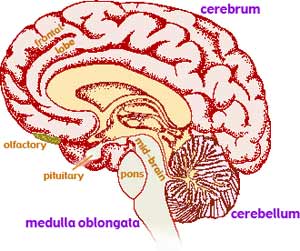
The cerebrum is the largest region of the mammalian brain. It is typically folded, with species with more advanced brains having more folds. Presumably the folding helps distinguish which neurons share information. Different analysis processes are located in different regions of the cerebrum. For example, many animals have a major structure hanging down from the cerebrum into the nasal cavity. This olfactory bulb pre-processes odor information. In humans and other primates it is relatively small compared to more primitive creatures which are more dependent on their sense of smell for survival. Another small region near the front of the left brain trigger the feeling of mirth so when it is stimulated, a person to find humor in whatever they are seeing. The cerebrum consists of two nearly independent halves with very limited connects between the left and right halves. It has been suggested that the right half does the equivalent of parallel processing of information, providing a holistic impression of our world including ourselves. The left half may do the equivalent of serial processing of data, providing for analysis, pattern recognition, and language.
The brain undergoes transitions from consciously awake to asleep. Sleep and dreaming may provide a mechanism to consolidate gathered data, perhaps similar to restoration processes conducted periodically by computer operating systems. Much of what is known about the brain came from people who have damage to particular parts of their brains. One research method applies via very thin needles small electrical signals near damaged parts of the brain with the person reacting or reporting perceived changes. Other efforts have mapped the blood flow (which increases with metabolic rate 1 to 5 seconds after brain activity) when people are provided different visions or sensations or asked to provide particular thoughts or behaviors. But much remains to be learned about brain function.
The brain processes such as analysis and response to incoming information from the senses, and memory of that information seems to be accomplished between neurons at the junctions termed synapse.
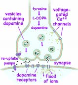
At the terminal end of the neuron's axon, the action potential (spike in Voltage) triggers the opening of channels which permit Calcium ions, Ca+2, to enter the neuron. The Calcium ions cause the secretion of neurotransmitters molecules from lipid-membraned capsules loaded and waiting just inside the axon. Once released into the synapse these neurotransmitters wander across the gap to receptors on the adjacent target neuron. 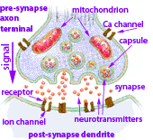 When the neurotransmitters bond to matching receptors they trigger the release of secondary messenger molecules inside the target neuron. These secondary messengers activate protein kinases (molecules which bind phosphate groups to other proteins). Some of the secondary messengers open additional channels in the cell membrane allowing Ca+2 to flow in and K+ ions to escape changing the electrical potential (Voltage across) the outer membrane of the target neuron, sending the electrical signal onward and triggering other changes. Finally the neurotransmitters are reabsorbed or deactivated and the synapse is prepared for handling another signal.
Glutamate and GABA, are the primary neurotransmitter molecules found in the brain. Over half of all brain synapses release glutamate, and 30 to 40% of all brain synapses release GABA. Glutamate serves to excite the receiving neuron while GABA inhibits the response of the receiving neuron. Both neurotransmitters work together to control many processes, including the brain's overall level of excitation. Other neurotransmitter molecules are described below.
In general, a strong action potential (spike in Voltage) travelling along one neuron will release enough neurotransmitters across the synapse to trigger a response continuing along the target neuron.  Whereas a weak incoming signal will not have enough action potential and as a result the signal will fade away. Each brain neuron can receives incoming signals from as many as a thousand other neurons via its dendrite synapses, and feed outgoing signals to a similar number of other neurons across its axon synapses. When action potentials fire nearly simultaneously in several neurons that weakly synapse on a single neuron, by summation they may initiate a signal impulse which has sufficient action potential to continue though the maze of neurons. Some synapse provide glutamate which excites their target. Other synapse provide GABA which inhibits their target, reducing the action potential and decreasing the likelihood of the signal continuing. Thus the output of a neuron depends on the input of many others, each have a different degree of influence. John Carew Eccles (b.1903, d.1997 Nobel photo at right →) received the Nobel Prize for Physiology/Medicine in 1963 for performed early experiments on such integration of synapse signals.
The brain has a series of interconnected neurons along which signals are transmitted from one brain region to another. Dopamine, and serotonin are neurotransmitters which are produced by only a few neurons in the brainstem but distributed through their long axons to thousands of other neurons, thus having considerable influence over local brain processes. Dopamine seems to create a wanting or craving of that which was previously valuable or beneficial for the individual (such as food). It may serve the function of drawing our attention to nerve signals which may be of more significance. Dopamine is distributed by the mesolimbic pathway and also distributed by the nigrostriatal pathway and the tuberoinfundibular pathway. Serotonin is produced by neurons in the Raphe nuclei in the brain stem and is distributed to most of the brain and spinal cord. Serotonin plays a role in many regulative processes such as body temperature, sleep, mood and appetite.

Arvid Carlsson (b1923, ←Nobel photo at left) was awarded 1/3 of the 2000 Nobel Prize for Physiology/Medicine for discovering in the late 1950s that dopamine is a neurotransmitter in mammal brains. By administering reserpine to animals, he was able to reduce available dopamine creating the symptoms of Parkinson's disease which is a palsy characterized by muscle rigidity, tremor, a slowing of physical movement to complete loss of physical movement in extreme cases including impairment of speech and swallowing. Dopamine cannot be administered as a drug because it can't pass from the blood into the brain. However it is manufactured in the substantia nigra near the top of the brain stem from the amino acid tyrosine via the intermediate L-DOPA and then distributed to other neurons which repackage dopamine as a neurotransmitter. By then administering L-DOPA, Carlsson was able to restore normal brain function. This led to somewhat successful treatments for humans with Parkinson's disease. Dopamine has been the most intensively studied neurotransmitter because dopamine signal transmission difficulties underlies several major disorders:
Glutamate is a key molecule in cellular metabolism. It is one of the amino acids which, while abundant in the food we eat, our cells can also manufacture. α-ketoglutarate is an intermediate in the citric acid cycle which cells use to metabolize carbohydrates, both producing energy carrying ATP and GTP and allowing intermediaries to be syphoned off to produce other useful molecules. The amine group (–NH2) from another amino acid can be substituted for the ketone group (C=O) on the α-ketoglutarate to produce glutamate. Glutamic acid becomes the glutamate ion whenever a weakly acidic carboxyl group looses its Hydrogen ion (–COOH ↔ –COO-1 + H+1). With one of the Hydrogen ions replaced by a Sodium ion, it becomes monosodium glutamate, MSG. Glutamate and the closely related GABA both exist most of the time in aqueous solutions as zwitterions with the Hydrogen ion, H+1, departed from the –COOH leaving it a negatively charged –COO-1, and the amine, –NH2, possessing an extra Hydrogen ion making it the positively charged –NH3+1.
Glutamate was discovered and identified by the German chemist Karl Heinrich Leopold Ritthausenin in 1866. In 1907 Kikunae Ikeda at Tokyo Imperial University identified brown crystals left behind after the evaporation of a large amount of kombu broth as glutamic acid. When tasted the crystals, he noted a flavor common to many foods, most especially in seaweed. Ikeda termed this flavor umami, one of the five basic tastes humans have receptors for on our taste buds. Glutamic acid is present in a wide variety of foods, including cheese and soy sauce. It is part of larger proteins found in all meats, poultry, fish, eggs, dairy products, as well as kombu. Ikeda patented his method of mass-producing the crystalline salt of glutamic acid, monosodium glutamate (MSG). Roughly two million tons of MSG is commonly used annually as a food additive and flavor enhancer, most heavily in China. Ninety-five percent of the dietary glutamate is metabolized in the intestine and very little passes from the blood into the brain. So glutamate used as a neurotransmitter is essentially manufactured as needed in the brain.
For use at synapses between nerves, glutamate is stored in vesicles inside the axon terminals. A voltage spike travelling down the axon causes the lipid membranes encasing the glutamate to merge with the cell membrane, popping open and releasing the glutamate into the synaptic gap. Those glutamate molecules diffuse across the narrow synapse where many bind to receptors on the neighboring dendrite surface triggering the opening of gated channels allowing Calcium ions to enter that neuron. Any residual glutamate is rapidly removed from the space outside either neuron so that the synapse is prepared for arrival of another voltage spike. In invertebrates, particularly arthropods and nematodes, glutamate has a somewhat different effect, triggering the opening of gated chloride channels.
Failure to remove the glutamate from between the cells has been implicated in epileptic seizures.
Glutamic acid also serves as the precursor for the synthesis of the neurotransmitter GABA which when released, travels across the synapse and binds to receptors on both pre- and postsynaptic neurons. It inhibits the further response of both neurons.
| Neurotransmitter | Structure | Effects in Brain |
|---|---|---|
| Glutamic acid, (glutamate when ionized) |
It is the most abundant, swift, excitatory neurotransmitter in the mammalian nervous system. It is believed to be involved in cognitive functions like learning and memory. | |
| GABA (γ amino-butyric acid) |
An amino acid found only in the nervous system, it functions as the chief inhibitory neurotransmitter in the mature brain. It causes the opening of ion channels to allow the flow of either Chloride ions, Cl-1, into the cell or Potassium ions, K+1, out of the cell. This action results in charging the interior of the neuron negative. (In a brain prior to birth when glutamate synapses haven't yet matured, the GABA receptors cause the opposite flow of charged ions resulting in an excitatory effect.) | |
| Dopamine | Involved with endocrine & motor systems, also with cognition,
motivation, wanting, craving, desire, (but not the pleasure of enjoying an obtaining reward), and nausea Dopamine in the mesolimbic pathway increases general arousal and goal directed behaviors and decreases latent inhibition; all three effects increase the creative drive of idea generation. Novelty stimulates dopamine circuits in the brain, causing optimism and elation. |
|
| Serotonin | 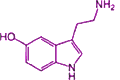 |
modulation of anger and aggression, body temperature, mood, and sleep. Increases introverted personality, sexuality, appetite, satiety, while decreases pain reception. |
| Norepinephrine |  |
synthesized from dopamine, affects large areas of the brain where attention and responding actions are controlled
|
| Acetylcholine | Acetylcholine is the only neurotransmitter used in the somatic nervous system where it causes the contraction of voluntary muscles; it is also used in the autonomic nervous system and in all autonomic ganglia.
|
|
| oxytocin → and vasopressin |
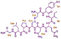 |
social recognition, bonding, generosity and the formation of trust. It initiates and supports pair-bond between mates |
There are two distinct mechanisms, distinguished as fast and slow synaptic transmission, by which nerve cells communicate with their neighbor cells.
Rodolfo Linas showed fast-acting neurotransmitters, e.g., glutamate (which excites) and GABA (which inhibits), effect their neighbor cells within one millisecond. About half of the brain's fast synapses excite a target neuron, mostly with glutamate. The other half of the fast synapses inhibit their target neuron, mostly using GABA. The fast mechanism involves neurotransmitters attaching to a receptor which is directly connect to an ion channel which quickly snaps open causing an immediate flood of ions.
For example, a glutamate binds to a receptor with a closed gate causing the gate controlled by this receptor to change shape, opening to allowing Sodium ions, Na+1, to flood across the membrane. The positively charged Na+1 cause a voltage reversal across this membrane which helps create a Voltage spike in the receiving nerve.
If instead of glutamate the neurotransmitter is GABA, the resulting flood of ions are not Sodium but chloride ions. The negatively charged chloride ions, Cl-1 increases the membrane's negative Voltage thereby inhibiting any response by the nerve. The combination of exciting and inhibiting is used to establish memory and provide plèasure as explained further in the following two investigations, Biochemistry 8 and Biochemistry 10.
The slow synapse transmission is much more complex. All of the amine and protein neurotransmitters produce their effects on their target cells by slow mechanisms. Even the fast acting neurotransmitters, glutamate and GABA, also produce effects using slow mechanisms. Slow synaptic transmission are achieved over hundreds of milliseconds to minutes. This process is mediated through a complicated sequence of biochemical steps, involving second messengers, protein kinases, and protein phosphatases.
In the slow mechanisms, the neurotransmitters activate receptors causing a series of chemical reactions. First, one of several secondary messengers is produced. These could be cyclic AMP, cyclic GMP, calcium ions or diisoglycerol. These second messengers in turn activated messenger specific protein kinases. Angus Naim and Ivar Walaas found that these protein kinases add phosphate to more than 100 different substrate proteins in the brain which fall into four groups which in turn control the fast transmissions:
gainof the system. It determines whether a neurotransmitter molecule will be effective or not in activating a receptor by its state of phosphorylation.
 A group led by Paul Greenard (b 1925, ←Nobel photo at left) was inspired by studies that were carried out by Sutherland and Krebs who had been studying the mechanism by which the hormones epinephrine and glucagon break down glycogen in liver. Earl Sutherland found that epinephrine and glucagon cause the activation of a protein enzyme which caused the conversion of ATP to cyclicAMP. The cyclicAMP could mimic the hormones, causing the breakdown of the glycogen. Edwin Krebs found that cyclicAMP broke down the glycogen by activating a protein kinase enzyme. Protein kinases transfer phosphate from adenosine triphosphate (ATP) to to other molecules leaving adenosine diphosphate (ADP). Krebs and his colleagues found that the protein receiving the phosphate was a phosphorylase kinase enzyme which in turn catalyzed the breakdown of the glycogen. Another enzyme deactivated the phosphorylase kinase enzyme by removing the phosphate. Greenard wondered if a similar process was used at the synapse. They found in the nervous system a family of protein kinases that were activated by neurotransmitters, including enzymes sensitive to the amines dopamine and serotonin. They found some other second messenger-dependent protein kinases about the same time as they found a protein kinase which got activated by cyclicGMP rather than cyclicAMP. CyclicGMP is a short term for guanosine monophosphate which has a multiple-ring shape. Later, they found a protein kinase that was stimulated by Calcium.
They found a family of proteins which they called synapsins. The extent to which these synapsin proteins have attached phosphate groups modulates the release of neurotransmitters across the synapse. Synapsin I without phosphate functions to tether vesicles containing the neurotransmitters in a reserve
pool. When phosphate is attached to synapsin I, this tethering ends, and the vesicles are then free to respond when a signal comes down the surface of the nerve Greengard was awarded 1/3 of the 2000 Nobel Prize for Physiology/Medicine for showing what happens when dopamine and similar transmitters stimulate a nerve cell. Receptors on the cell surface activate enzymes in the cell membrane, which starts the production of second messengers. These messengers travel into the cell and activate a protein kinase, which starts to bind phosphate groups to other proteins, in this way altering their function. This leads, for example, to the opening of ion channels in the cell membrane and a change in the electrical activity of the cell.
Greengard's group found a compound, DARPP-32 (an acronym for dopamine and cyclic AMP regulated phospho-protein of 32,000 Dalton molecular mass). Dopamine and about a dozen other neurotransmitters which regulate the activity of these neurons do so in large measure by regulating the state of phosphorylation of a pivotal signaling switch referred to as DARPP-32. The state of phosphorylation of DARPP-32, through control of protein kinase and phosphatase activities, regulates the state of phosphorylation and activity of virtually every known physiological effector, including a variety of ion pumps, ion channels, neurotransmitter receptors and transcription factors. The essential role of DARPP-32 in cell signaling in the brain extends far beyond the dopamine cell-signaling system and appears to involve a large number of neurotransmitters in many brain regions. The phosphorylated form, but not the dephosphorylated form, of DARPP-32 inhibits a protein phosphatase which in turn controls the activity of various ion pumps and channels. Thus, the actions of numerous neurotransmitters in producing physiological effects in these neurons can be accounted for in terms of a complex signal transduction cascade. The DARPP-32 is a bifunctional compound which can be either a kinase inhibitor or a phophatase inhibitor, depending on the information coming into these cells.
As is often the case in science, new discoveries often follow the development of a new tool or technology. In the case of brain mapping, the new tool is the MRI, Magnetic Resonance Imaging, which uses the unique magnets inside particular atomic nuclei which have unpaired magnetic fields due to the odd number of protons and neutrons such as 11H, 178O, 15564Gd and15764Gd. These are excited to resonate by tuning strong radio frequency vibrations to match the natural frequency of the particular kind of atom being used. The measurements are done in a very strong, necessarily very stable which have well measured magnetic fields. When the radio waves are briefly turned off, the excited atomic nuclei align with the very intense magnetic fields slightly warping the fields allowing their presence can be detected by mapping the changes in the field. A secondary gradient magnetic field is applied by additional magnets so that only a single plane in the field will precisely tuned to receive energy from the excited atomic nuclei. By changing the magnetic gradient and repeating the measurements, a series of flat images can be gathered allowing the 3-D reconstruction of where the chosen atomic nuclei were located. 11H is most often used because of its availability in water and the different water concentrations in different tissues. But for brain activity to be mapped by metabolism changes, 178O is often used because active neurons require additional Oxygen carried by hemoglobin in blood.
![]()
to next investigation
to Biochemistry menu
to ie-Chemistry menu
to site menu